Enzyme metallography is also a highly effective method for the fabrication of conductive array biochips.
DNA sequences are detected by the formation of conductive electrical contacts, between pairs of electrodes linked by printed dots of complementary capture oligos. Once the targets hybridize to the capture oligos, the targets are detected using a haptenated probe which is in turn bound by a streptavidin-peroxidase polymer which is then developed using EnzMet™.
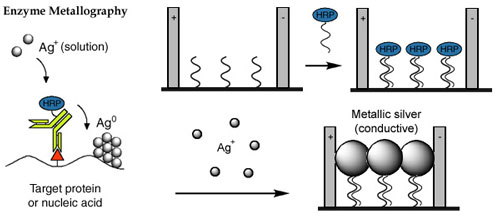
The process of creating a conductive array biochip with EnzMet™ was demonstrated using labeled targets:
The biochip developed using EnzMet™ was compared with an equivalent biochip, in which detection was achieved using silver-enhanced gold labels.
Examination of the two biochips using atomic force microscopy (AFM) revealed a significant reduction in background signal using EnzMet™, with fewer silver deposits in the substrate areas adjacent to the spotted region. A highly defined separation was found between the spot region with deposited metal of about 130 nm height and the surrounding background, with no detectable metal clusters.
Furthermore, the electrode regions that are situated inside the spot area also show a clearly suppressed metal deposition with the enzymatic process compared with images from the metal-catalyzed case. This lower background signal enables the enzyme-based detection scheme to be significantly more sensitive than the nanoparticle-based system: it was found to be possible to detect biotin-modified target DNA concentrations as low as 500 fM, corresponding to 50 amol of biotin-modified target DNA.
Because multiple independent electrical contacts can be fabricated and identified on a microscopic scale, electrical detection offers the potential for highly multiplexed target detection in a robust, miniaturized and highly portable format.
Previous studies had demonstrated that using silver-enhanced gold nanoparticles could be used to form the contacts, but produced significant background staining even after treatment with passivating agents such as alkanethiols or mercaptoalcohols.
Because of its high sensitivity and low background, enzyme metallography offers the potential for improved detection.
For biochip fabrication, order
EnzMet™ for General Research Applications.
Moller, R.; Powell, R. D.; Hainfeld, J. F., and Fritzsche, W.: Enzymatic control of metal deposition as key step for a low-background electrical detection for DNA chips. Nano Lett., 5, 1475-1482 (2005).